


A number of assessment tools can be used to determine if a patient is at risk of myocardial damage caused by cardiometabolic disease and/or COVID infection. Many of these tests also reflect the general dysfunction of cellular metabolism that underlies a host of chronic conditions.
Vitamin D3 Level: There are so many good reasons to measure a patient’s level of 25-hydroxyvitamin D (25-OHD) in the blood. Vitamin D3 is important for the prevention and treatment of heart disease and viral infections. It is a simple inverse relationship: if vitamin D is low, risk is higher. Statistically, low vitamin D correlates strongly with COVID risk. Vitamin D also activates many cancer prevention genes; women with high levels of vitamin D3 (60-90 ng/ml) have an 82% lower risk for breast cancer.
Most studies indicate that risk of CVD rises when vitamin D levels drop below 30 ng/ml. Optimal levels for protection against CVD are in the range of 60-90 ng/ml.
GlycoMark: The GlycoMark test measures blood levels of 1,5-Anhydroglucitol (1,5-AG), a glucose-like sugar found in many foods. It is an indicator of the average maximum blood glucose level over the previous 14 days. A sharp decrease in circulating 1,5-AG signals that a patient has frequent post-prandial glucose surges and greater risk.
According to the company that makes the test, two people with similar A1C levels in the 7-8% range may have very different glucose variations throughout a day. Roughly 40% of people with “healthy” A1Cs have persistent glucose spikes, putting them at high risk for cardiometabolic complications.
GlycoMark is an adjunct to standard glucose tests. It is a simple well-validated tool for early detection of glucose dysregulation. A reading of 3 ug/ml or lower indicates potential problems; a reading above 10 indicates better glucose regulation.
Homocysteine: This is a great test to determine if the body has adequate levels of B vitamins, especially B6, B12 and folic acid. Deficiencies in these vitamins often indicate future cardiovascular and cognitive problems, as well as hormone-related diseases, potential birth defects, and many other health challenges.
C-Reactive Protein (CRP): Inflammation is a key driver of CVD, along with other chronic diseases and CRP is a reliable indicator of inflammation. It provides an early warning for heart disease and many other chronic problems. Often triggered by the presence of free radicals and oxidative stress, high CRP is an early warning sign for heart disease, cancer, and other illnesses.
8-OHgD test: 8-hydroxy- 2’-deoxyguanosine (8-OHdG) is a byproduct of oxidative damage to guanine bases in DNA, which makes it a good indicator of DNA damage caused by chemical toxins in the air, water or food, as well as various forms of radiation. A high level of 8-OHgD in urine indicates increased oxidative stress, thus providing an early warning of disease. The acceptable or “normal” levels on the 8-OHgD urine test are 43.9+/-42.1 ng/mg creatinine in females, and 29.6+/-24.5 ng/mg creatinine in males.
Galectin-3 (Gal-3): This molecule plays important roles in normal cell adhesion, cell growth, and chemoattraction. However, Gal-3 elevations indicates that something is wrong. High Gal-3 is strongly associated with ventricular remodeling, heart failure, and stroke. It is also associated with some forms of cancer. Gal-3 is over-expressed on cancer cell surfaces, and actually helps them to stick to one another. It also circulates in the blood, indicating that cancers are attempting to metastasize.
Because it can signal the possibility of multiple diseases, Gal-3 measurement is not a stand-alone diagnostic test for any particular condition. But it is a useful biomarker of chronic inflammation, tissue fibrosis, and possible neoplasia. From a CVD perspective, serum Gal-3 levels below 18 ng/ml are considered low-risk; readings above 25 ng/ml suggest high risk for heart failure.
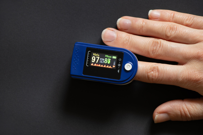
Oximetry: A simple finger oximeter can provide important signals. If oxygen levels are low, the mitochondria do not produce enough energy and cells are not able to function properly. Most finger oximeters use a 100-point scale: the desired level is from 96 to 100. As oxygen level drops, cellular metabolic problems increase. Vascular damage and vasoconstriction are primary causes of low readings. In some cases, this reflects infection with SARS-CoV-2.
Vascular Endothelial Growth Factor (VEGF): VEGF is a signal protein that stimulates the growth of new blood vessels in response to low levels of oxygen. It can also produce a profound increase in vascular permeability and edema. Over- and under-expression of VEGF can both create problems. Too much VEGF can lead to excess vessel growth and excess permeability of capillaries. Too little can lead to a lack of oxygenation in tissues, which can trigger symptoms of chronic inflammatory response syndrome (CIRS).
Most studies establish the optimal range of VEGF to be between 31 pg/ml and 86 pg/ml.
Gamma-Glutamyltransferase (GGT): This enzyme is found in many organs in the body, with higher concentrations in the liver where it helps to deliver cysteine for intercellular glutathione synthesis. While predominately a marker of liver disease, elevated serum GGT also indicates oxidative stress, often associated with excessive alcohol consumption, heavy metal exposure, heart disease, and diabetes. High GGT can also be associated with increased cancer risk. Typical reference ranges are 15-85 IU/L for men, and 5-55 IU/L for women.
Magnesium RBC: Magnesium has many physiological roles. One of the most important is glucose and insulin transport. The level of magnesium in red blood cells is the best indicator of overall magnesium levels in the body. Mg deficiency is associated with diabetes, and a host of cardiometabolic problems. It is also linked to increased cancer risk. The reference range for magnesium RBC tests is 4.2−6.8 mg/dL.
D-dimer: D-dimer is a small protein fragment that results from fibrinolysis. High levels are associated with deep vein thrombosis, pulmonary embolism, and stroke. It is also a biomarker of abnormal clots associated with COVID-19 infection or in response to a COVID vaccine (if the test is done within ten days of diagnosis or injection). A four-fold increase in D-dimer is an indicator of poor prognosis in hospitalized COVID patients. In adult males and non-pregnant females, the threshold is 0.5 ug/ml.
Omega 3/6 ratio: This test measures the ratio of omega-3 to omega-6 fatty acids in the blood. In broad-stroke terms, omega-3 is anti-inflammatory, while omega 6 is inflammatory. We need both to maintain health, but they need to be in the right balance. Too much omega-6 drives inflammation and raises risk of CVD and other diseases. A good ratio is in the range of 2 to1 or 3 to1, favoring the omega-3s.
MRI Plaque View: This relatively new assessment tool shows the actual morphology of atherosclerotic plaques, including multi-contrast identification of the lipid core, calcification, and hemorrhage potential. It indicates maximum carotid wall thickness, normalized wall index, and gives a lot of information about the health of the endothelial lining. It is an accurate way to assess plaque vulnerability to rupture, thus giving a clear picture of thrombosis risk.
PULS Cardiac Test: The Protein Unstable Lesion Signature (PULS) Cardiac Test is another relatively new method that aggregates biomarkers of immune system activation and endothelial injury, including IL-16, MCP-3, Eotaxin, and sFas, along with HDL and hemoglobin A1c. It combines these variables into a composite score that predicts the odds that someone will have a heart attack within five years.
There is good science to support this method, as summarized in a recent paper by Premyodhin and colleagues in Coronary Artery Disease. But PULS testing is not yet considered standard of care in cardiology.
Cardiac surgeon Steven Gundry recently published an abstract looking at the effect of mRNA COVID vaccines on PULS scores. Gundry has been using PULS regularly for 8 years. He ran the PULS tests 2-10 weeks following patients’ second doses of mRNA shots, and compared the scores to each patient’s pre-vaccine levels.
Based on those comparisons, he observed increases in IL-16, sFas, and HGF (hepatocyte growth factor), which raised 5-year coronary event risk scores from an average 11% to 25%. Gundry concludes that mRNA vaccines trigger endothelial inflammation.
The findings are not entirely surprising given that the purpose of a vaccine is to generate temporary inflammation. The question is whether the observed elevations are dangerous.
Gundry’s experiment did not include an unvaccinated control group, patients who received a non-mRNA vaccine, or people receiving any other type of conventional non-COVID vaccine. The paper gives no data about duration of the inflammatory surges.
Further, there is no information given about PULS score changes during an acute COVID episode, which makes it difficult to gauge the potential vaccine risk Gundry is suggesting against the risks associated with severe COVID. His observations raise important questions that warrant attention. But without these comparators they are not conclusive.
Charles K. Bens, PhD, is a nutritionist and educator based in Sarasota, FL. He is the founder of Healthy at Work, a corporate wellness program, and the author of 9 books, including Healthy at Work: Health in Your Pocket: Your Pocket Guide to Good Health. Reach Dr. Bens at: bensck@gmail.com
END