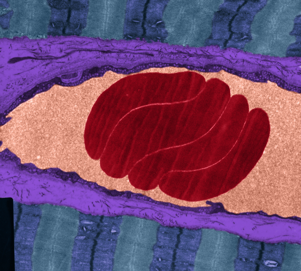
(Image: OMIKRON/Science Photo Library)
Repair of the endothelial glycocalyx is a vital element in the restoration of cardiovascular health, according to Mark Houston, MD, director of the Hypertension Institute at Saint Thomas West Hospital, Nashville.
The endothelial glycocalyx (EGX) —literally “sugar coating” in Greek—is a very thin gel-like layer that lines the luminal surface of the blood vessels. It is a carbohydrate-rich mesh of membrane-bound and soluble glycoproteins, proteoglycans, and glycosaminoglycans, which create a non-adherent shield.
Damage to this sensitive, bioactive layer is one of the earliest steps in the pathogenesis of cardiovascular disease, Dr. Houston said at the 2022 Integrative Healthcare Symposium.
“Endothelial dysfunction is the starting point of CVD, and the EGX is the primary protector of the endothelium. Therefore, maintaining a healthy EGX may be one of the most important intervention targets for prevention of CVD.”
A “SMART” Barrier
To understand the importance of the EGX keep in mind that the vascular endothelium is not passive. It actively controls passage of nutrients, oxygen, and hormones into and out of the bloodstream. It is also a key player in immunity and inflammation. Through production of nitric oxide, it regulates blood pressure.
“The endothelium affects the structure and function of every other organ system,” Houston explained. “Whatever the glycocalyx does, the endothelium does as well.”
According to Dr. Houston, the glycocalyx is a “SMART” barrier:
Selective: It is selectively permeable, preventing cholesterol, platelets, leukocytes, and other circulating blood components from sticking to vessel walls, but allowing passage of nutrients, dissolved gases, and signaling molecules.
Micro-thin: The thickness of the EGX varies with the thickness of the arteries it coats. It is generally in the range of 2-3 um in small vessels, and up to 4.5 um in larger ones like the carotids. Even at its thickest, it is still quite thin. It would take roughly 1,000 layers of EGX to equal the thickness of one sheet of paper.
Antioxidant: The EGX harbors the antioxidative enzyme, superoxide dismutase (SOD), which reduces oxidative stress, and keeps nitric oxide (NO) available to the vasculature.
Regulator: It regulates vascular permeability, inflammation, coagulation, and fluid balance.
Transducer: The EGX contains brush-like structures in the 150-400 nm range, comprised of glycoprotein and proteoglycan chains. These sense the shear stress of blood flow, and signal the endothelium to produce nitric oxide when needed to regulate vascular tone. It also responds to chemokines, cytokines, and other molecular signals involved in vascular homeostasis.

One of its primary functions is to protect the endothelium from thrombus formation. It blocks the binding of sticky leukocytes, activated platelets, and lipoproteins–especially LDL. It also contains anti-thrombin III, tissue factor pathway inhibitors, lipoprotein lipase, vascular endothelial growth factor, superoxide dismutase, and hyaluronic acid, all of which mitigate inflammation and resist thrombus formation.
Negatively charged glycosaminoglycans within the EGX bind and inactivate sodium, rendering it non-osmotic and preventing it from accumulating in the endothelium. Thus, the EGX buffers against salt-induced arterial stiffness.
Hyperglycemia & EGX Damage
Damage to the EGX is a direct consequence of persistent hyperglycemia, dyslipidemia, chronic inflammation, and oxidative stress. It precedes all the vascular complications of diabetes.
“Endothelial dysfunction is the starting point of CVD, and the EGX is the primary protector of the endothelium. Therefore, maintaining a healthy EGX may be one of the most important intervention targets for prevention of CVD.”
Mark Houston, MD, Director, Hypertension Institute at Saint Thomas West Hospital, Nashville
“High blood glucose causes damage to glycocalyx, even in the so-called “normal” range. You want fasting blood sugars down to 75, and the A1c down to 5. Anything above that will increase risk because that’s when the glycocalyx damage starts,” Houston told IHS attendees.
Many other things also contribute to glycocalyx degradation, including elevated TNF-a, hypervolemia, low fluid sheer stress, elevated hyaluronidase, matrix metalloproteinases, atrial natriuretic peptide, and bacterial endotoxins.
There’s a clear and simple correlation between reduced EGX thickness and predisposition to lesion formation. Houston noted that in “atheroprone” regions of the vasculature–such as vessel branches, bifurcations, and curvatures—the EGX is typically very thin.
Restoring the Glycocalyx
A number of treatments and factors can help to restore, regenerate, or maintain a healthy EGX, including:
- Hydrocortisone
- Calcium channel blockers
- Glycemic control via Metformin
- Sulodexide—a highly purified mixture of glycosaminoglycans composed of low molecular weight heparin (80%) and dermatan sulfate (20%)
- Statins
- Reduction of inflammatory mediators
- Nitric oxide
- Albumin
- Fresh frozen plasma (FFP)
- N-acetyl cysteine (NAC)
- Hydroxyethyl starch
- Sphingosine-1 phosphate (SIP)
- Glycocalyx-regenerating compounds (GRCs): hyaluronan, antithrombin III, heparin, sulodexide, specialized sulfated polysaccharides (SSP), and protein C
But Dr. Houston holds that the single most effective and convenient option is Rhamnan sulfate (RS), a sulfated polysaccharide extracted from two types of marine algae (Monostroma Latissium and Monostroma Nitidum). These polysaccharides
have similar structure to glycosaminoglycans found in the human EGX.
Rhamnan Sulfate: Safe & Effective
Early in vitro experiments showed clearly that rhamnan sulfate can repair EGX damage caused by excessive glucose exposure. Animal experiments show that it prevents leukocyte adhesion, suggesting that it might prevent endothelial inflammation at its earliest stages.
Rhamnan sulfate is available as a dietary supplement called Arterosil. This product also contains grape seed extract, green tea extract, and concentrated extracts from a host of heart-healthy fruits and vegetables.
Supplementation with rhamnan sulfate can markedly increase arterial elasticity, a good indicator of endothelial function. In a study of 19 healthy humans at Baylor College of Medicine, daily supplementation with Arterosil increased carotid arterial elasticity by an average of 89.6% over baseline, within two hours of ingestion.
“Arteries are arteries. If it does this in the carotids, my guess is that it does it everywhere,” said Dr. Houston. “If you give a glycocalyx promotor, you can get increases of vascular compliance very quickly, and the functional changes beget structural changes.” He added that he is now recommending Arterosil and other glycocalyx-promoting treatments to 100% of the patients in his clinic.
“The average increase in arterial elasticity is almost 90%, very significant improvements.”
Plaque Reversal
Houston contends that conventional medicine’s myopic obsession with lipids has blinded many physicians to the myriad other factors that drive vascular pathology.
“Even if you use statins and drive down LDL to 40, still 50% of patients will have events. Lipids are not the only problem in CAD. There are many other steps.”
That said, new evidence indicates that rhamnan sulfate can reduce atherosclerotic plaque formation, at least in mice. Notably, the plaque-reducing and anti-inflammatory effects were stronger in female versus male animals.
Dr. Houston reported early data from an ongoing human pilot study of rhamnan sulfate in patients with vulnerable atherosclerotic plaques, as confirmed by MRI-PlaqueView imaging. PlaqueView is the only FDA-approved software system for carotid plaque analysis.
He pointed out that stenotic plaques account for less than 50% of all “culprit” lesions, whereas 75% of all events are attributable to ruptured non-stenotic plaques that trigger thrombus formation.
Daily supplementation with a rhamnan sulfate product for two months led to a mean 64% regression of lipid-rich necrotic plaque—the most dangerous type of plaque—in the female participants, and a 47% regression in the males. There were also significant increases in vessel lumen diameter, suggesting a reduction of carotid stenosis—a finding that has not been observed in statin studies.
“Rhamnan sulfate changes the morphology of plaques, making them more stable. These are very significant changes. Nothing else does this. No drug can do this. Statins reduce lipid-rich necrotic plaques by about 25%, which is meaningful, but not even close to what you get with the Rhamnan sulfate.”
These preliminary findings from China, have prompted a larger US-based study which is now underway.
Houston stressed that plaque reversal is a gradual process. Typically, it takes about 6 months of twice-daily dosing to see reversal of carotid plaques. “For really bad cases, you can take two caps, twice daily. The key is to split the doses, ideally 12 hours apart. It works better that way.”
“Rhamnan sulfate changes the morphology of plaques, making them more stable. These are very significant changes. Nothing else does this. No drug can do this. Statins reduce lipid-rich necrotic plaques by about 25%, which is meaningful, but not even close to what you get with the Rhamnan sulfate.”
Mark Houston, MD, Director, Hypertension Institute at Saint Thomas West Hospital, Nashville
Rhamnan sulfate does not break up plaques and cause fragments to float away. Rather it promotes resorption of the lipid core while simultaneously blocking deposition of lipids. It does this without actually affecting serum lipid levels.
According to Dr. Houston, Aterosil is safe, and unlikely to interact adversely with medications. In fact, it is likely to be synergistic with Rosuvastatin (Crestor) and other statins. “There are zero drug interactions with this. In combination with nearly everything we use, the interactions are generally good. If anything, it makes the drugs more effective.”
Impact on Blood Pressure
Houston and his colleagues at the St. Thomas West Hospital studied Arterosil in a cohort of ten patients with uncontrolled hypertension. After three months of twice-daily dosing, the patients showed a decrease in mean systolic pressure from 151.5 mmHg at baseline, down to 147.5 at the three-month point. Diastolic pressure dropped from a baseline mean of 93.2 down to 82.3 mmHg.
His team is currently looking at the impact of this product on diabetic neuropathy in a cohort of 20 patients. He says “preliminary results are very promising.”
Potential Role in Covid Care
The glycocalyx and its restoration has implications in the context of Covid-19.
A healthy EGX may reduce susceptibility to viral infection, as well as the risk of severe Covid symptoms if one does get infected. Conversely, the Covid disease process can damage the glycocalyx, Dr. Houston explained.
Early on in the pandemic, investigators at the Lawson Health Research Institute, Ontario, reported that ICU patients infected with the then-novel virus showed marked glycocalyx degradation compared with ICU patients who were virus-negative. This correlated with increases in thrombosis, depressed nitric oxide production, and increased platelet adhesion (Fraser DD, et al. Crit Care Explor. 2020).
“A therapeutic strategy based on glycocalyx protection would be effective for Covid-19 patients with both early and severe (e.g., ARDS) disease.”
Hideshi Okada, MD, Gifu University, Japan
A team at the Jagiellonian University, Krakow, Poland showed that in human pulmonary arteries, the intact EGX strongly binds viral spike protein, “but screens its interaction with ACE2.” When the glycocalyx is reduced, ACE2 receptors on the surface of the endothelial cells are exposed, enabling the spike proteins to bind. The Krakow group concluded that susceptibility to Covid-19 may depend on the condition of the glycocalyx.
A Greek research team showed strong correlations between reduced EGX thickness, oxidative stress, vascular dysfunction, and impaired cardiac performance in patients with SARS-CoV-2.
Dr. Houston noted that “even in mild cases of COVID-19, EGX damage can persist for up to four months, and has been correlated with a 10-fold elevation in oxidative stress compared to controls. If the ACE2 enzyme is low to begin with, something like Covid, which depletes ACE2, will cause big, big problems. This is what happens in very bad cases.”
It is notable that people on ACE2-sparing drugs tend to have milder Covid cases. “You can increase the effects of ACE2 with a glycocalyx promoter,” Houston says.
In their excellent review of vascular injury in Covid-19, Dr. Hideshi Okada and colleagues at the Gifu University, Japan, point out that the endothelial glycocalyx is already very thin in pulmonary capillaries. Further degradation following an inflammatory cascade could be a major factor in acute and long-term Covid (Okada H, et al. Microcirculation. 2020).
“A therapeutic strategy based on glycocalyx protection would be effective for Covid-19 patients with both early and severe (e.g., ARDS) disease,” writes Okada. “A patient with comorbidity such as diabetes or hypertension would most likely exhibit an impaired glycocalyx function. Accordingly, their endothelial cells would not be fully protected and would be more susceptible to external (or internal) pathogens. In other words, the prevention of endothelial glycocalyx injury represents a useful means of systemic defense against infection.”
Blocking Viral Entry
Researchers at the Rensselaer Polytechnic Institute showed in cell culture experiments that rhamnan sulfate binds the spike protein binding domains on SARS-CoV-2, inhibiting interaction with ACE2 and heparan sulfate on endothelial cell surfaces. The effect of the seaweed extract was stronger than that of heparin or of pseudoviral particles being tested as potential Covid treatments.
The Rensselaer team reported strong antiviral activities against wild type SARS-CoV-2 as well as the delta variant (Song Y, et al. Mar Drugs. 2021).
Animal experiments at Chubu University in Japan suggest that rhamnan sulfate can directly inhibit influenza virus infection, while also promoting antibody production (Terasawa M, et al. Mar Drugs. 2020). They conclude that it is a potential candidate for the treatment of influenza virus infections
END


